
Laboratory of Advanced Technology
A Collaboration between Academia and Industry
Insights &
Updates
Interested in our services ?
Near-field optical microscopy … infrared spectroscopy at the nanoscale!
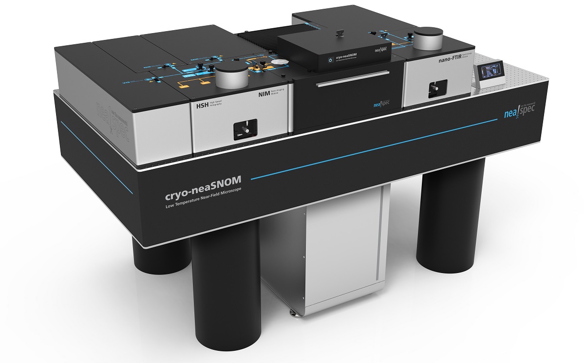
SNOM microscope now accessible through the LTA!
This neaSNOM microscope combines atomic force microscopy, optical imaging and nanoscale spectroscopy.
The capabilities of this Scanning Nearfield Optical Microscope (SNOM) include:
- Broad-band spectroscopy (terahertz-UV) with a spatial resolution of 10 mm, which is beyond the diffraction limit!
- Possibility to cool the sample at temperatures below 10K which is unique in Switzerland for a SNOM microscope. This allows for example the study of materials which are soft at ambient temperatures (biological or organic samples) or the study of phase transitions occurring at very low temperatures.
- High vacuum cell ensuring stability of analyzed samples
Thus, the neaSNOM microscope makes it possible to study the chemical, structural and electronic properties of a sample. The non-destructive measurement method is also suitable for organic and inorganic samples.
Examples of advanced nanoanalytical applications
Nanophotonics for optical information security
To ensure the authenticity or uniqueness of an object, it is possible to create a plasmonic structure at the nanoscale with a single near-field optical mapping undetectable by other methods.
Based on the principle of nanophotonics, it consists of using local interactions between nanometric and nanophotonic structures via near optical fields.
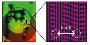
Schematic diagram of the nano / macro-hierarchical hologram concept with embedded nanophotonic code in the relief structure of the hologram.
Detailed analysis of stress fields around nanoindentation sites in silicon carbide (SiC) crystal
Near-infrared microscopy provides a non-destructive diagnostic tool for the study of local stress / strain fields and the propagation of nanofissures.
The structural modifications generating modifications of the electronic properties such as conductivity, can be detected and mapped with a nanometric resolution.
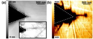
Zoom in the right corner of a triangular indent revealing a nanofissure emanating from the edge.
(a) Topography and (b) imaging s-SNOM
Nanoscale protein mapping: a new era in infrared nano-bio-spectroscopy
The structure of proteins determines their mechanical and catalytic properties and also plays a major role in many diseases. Near-field microscopy can address the challenge of recognizing and mapping the secondary structure of proteins with nanoscale spatial resolution.
The nanofocus at the top of the tip, which can be considered as a ultra-small infrared light source, allows sensitivity to unique protein complexes of less than one attogram (10-18 grams).
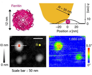
Nano-FTIR spectroscopy of individual protein complexes of ferritin